AMD
Age-related Macular Degeneration (AMD) is the leading cause of blindness in French patients older than 50.
It is estimated that more than a million French patients have AMD.
In a show "Capital Santé" (broadcasted via social networks, on the internet and on TNT), Pr Souied explains in detail what "AMD" means. It explains the main visual symptoms as well as depicting the two forms: atrophic and exudative. The risk factors and research perspectives are also discussed in this interview.
You can watch this show on Youtube.

Age-Related Maculopathy (ARM)
ARM represents an early stage of Age-related macular degeneration. It induces little or no visual symptoms.
It is characterized by the presence of alterations in the pigment epithelium and / or deposits in the retina called drusen in the macula (central area of the retina). If ARM is present in one or both eyes of a patient, his or her risk of developing AMD within 5 years is approximately 50%. Depending upon the extent of ARM, preventive treatments can be prescribed including antioxidants.

Age-related Macular Degeneration
By definition it is a disease of the macula that can appear from the age of 50 - 55 years. Two forms of Age-related Macular Degeneration exist: the atrophic form (or dry) and the exudative one (or wet).
Clinical signs
The clinical signs of AMD are metamorphopsia (the perception of wavy lines), and scotoma (the perception of a spot within the central visual field).


The atrophic form
It is the result of progressive atrophy of the pigment epithelium and photoreceptors. Impact on visual acuity is meaningful, but this process evolves in a relatively slow fashion.

The exsudative form
Twice as frequent as the dry form of the disease, the wet form is characterized by the appearance of abnormal choroidal neovascularization in the macula. These vessels are responsible for the formation of edema, intra- and sub-retinal exudates, and retinal hemorrhage. Wet AMD typically evolves and progresses rapidly.
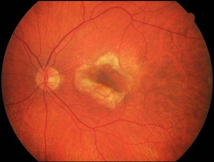
How is AMD diagnosed ?
Through a combination of fundoscopic examination and advanced retinal imaging:
These different tests are required to validate the diagnosis, initiate treatment, and ensure appropriate follow up care adapted to disease progression.


Which therapies are currently available?
Dry AMD
There is currently no treatment for atrophic AMD. However many therapeutic protocols do exist. If you are interested in these research protocols, the CHIC Ophthalmology team will be pleased to welcome you and answer to your questions. Many therapeutic research protocols are actually underway in our service.
Wet AMD
The standard treatment for exudative AMD is intravitreal injection therapy.
The injections are performed under local anaesthesia in aseptic conditions. One injection lasts a few seconds and is usually painless. A sand grain sensation may persist within 24 hours of injection. New and innovative treatments are currently under investigation. Our ophthalmology department is currently involved in many therapeutic protocols.
You can make an appointment with one of the Ophthalmologists of the department who provide specific care dedicated to this pathology:


Dr Alexandra MiereUniversity Lecturer – Staff Physician

Dr Francesca AmorosoStaff Physician


Dr Oudy SemounStaff Physician


Dr Mayer SrourPhysician


The care pathway
What is AMD? How to detect it? what is the treatment?
In this video, ophthalmogists of our department describe AMD care in Créteil. And patients speak about there desease experience, and talk about there clinical visits and treatments.
Video duration: 22 min 01 s (only in French).